BEVA Dental Chart
We have updated our Privacy Policy. You can find out more here.
BEVA Logo
Equine dentistry Equine dentistry

Dental charts

A dental chart should be used to record the findings of a dental examination.

Annotating dental charts
There is no standard protocol on how to fill in a dental chart though the general principles between different people are similar. This guidance will demonstrate some basic principles, common abbreviations and some grading systems from peer-reviewed literature.
Incisors

The occlusion of the incisors should be assessed and included on the diagram. An example of a diagonal bite is shown below.

Missing or lost incisors should be shaded out on the diagram and included in the narrative. Diastemata are commonly marked (D) and the degree of periodontitis included (see later).

Cheek teeth occlusal anatomy

Dental pulp anatomy is well described and pulp numbers are shown below.

When these pulps are exposed this is abbreviated to PE and annotated on the drawing and included in the narrative.

Dental overgrowths

The abbreviation OG is commonly used to describe overgrowth.

Overgrowths should be annotated both in diagram and in the text section. Whilst drawing overgrowths they should be above the surface of the tooth (Fig 3), whereas excessive wear should be taken away from the surface.

Dental fractures

A number of different configurations of dental fractures exist.

Figure 6. Lateral Sagittal Fracture

Parasagittal fractures through pulps no1 and no2 can be present in both maxillary and mandibular cheek teeth. The missing portions of these teeth are commonly shaded out (Fig 6).

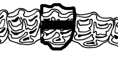
Midline sagittal fractures through the infundibula of the maxillary teeth are drawn similarly if a portion is missing. If both portions are present they are drawn as displacements with a black line in between over the infundibula (Fig 7).

Figure 7. Midline Sagittal Fracture with displacement

Occlusal fissure fractures are not commonly drawn on the diagram but are mentioned in the narrative.

A number of different configurations of dental fractures exist.

Infundibular caries

Caries may affect the cementum of the maxillary infundibula. A common abbreviation for infundibular caries is IC. It is common to mark the infundibulum affected on the occlusal portion of the chart with IC followed by the grade. An occlusal grading system is shown below (Fig 8). The depth of any lesion should be recorded in the narrative.

Figure 8. Infundibular Caries Grading System

Diastemata and periodontal disease

Figure 9. Diastema

As per the incisors diastemata are commonly annotated into an interdental space with an arrow (Fig 9).

Commonly they are abbreviated to D. Some classify these further into valve-type diastema D(V) or open-type D(O). Diastemata are commonly associated with periodontitis. This can be estimated as mild/moderate/severe or +/++/+++ (Fig 9). The depth of any periodontal pockets should be recorded.


Peripheral cemental caries

The cementum on the periphery of teeth may be affected by peripheral cemental caries. This is commonly abbreviated to PCC. A grading system is used and shown below (Fig 10). {Borkent:2016dw}

Figure 10